
When a patient is told that a liver tumour is “close to a major vein” or “near an important blood vessel,” it can be difficult to understand what that actually means from a standard scan.
For the surgeon, however, a few millimetres can make a considerable difference.
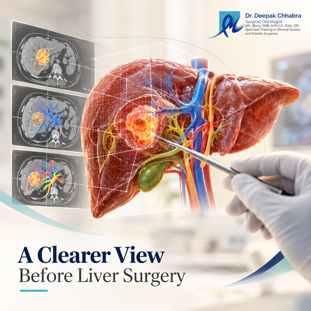
The liver contains a dense network of veins, arteries and bile ducts, and no two patients have exactly the same internal anatomy. 3D liver mapping takes information from CT or MRI scans and turns it into a patient-specific three-dimensional model, helping the surgical team see how a tumour sits among those structures before an operation is planned.
For patients exploring liver cancer treatment in Mumbai, understanding tumour location is only one part of the decision. The condition of the remaining liver, vascular anatomy and the amount of functioning liver that can safely remain also matter. Dr. Deepak Chhabra’s overview of liver cancer diagnosis and surgical treatment provides broader information about this treatment pathway.
3D liver mapping converts CT or MRI data into a model that shows the tumour in relation to the liver’s major veins, portal branches, arteries and—when imaging permits—bile ducts. It can help surgeons compare possible resection plans and estimate how much of the functioning liver is expected to remain after the tumour-bearing portion is removed.
Depending on the case, the model may help the team study:
The model is not a replacement for the original scan or the surgeon’s judgement. It is another way of seeing the same anatomy—often in a form that makes complex spatial relationships easier to understand.
Patients often focus first on the size written in the scan report.
A 3 cm tumour sounds easier than a 6 cm tumour.
Sometimes it is. Sometimes it is not.
A relatively small tumour positioned where several major vessels meet can create a more difficult surgical problem than a larger tumour sitting farther away from those structures.
Think of the liver less like a solid block and more like a city with roads running through it.
The tumour occupies one part of that city. The surgeon needs to remove the diseased area without unnecessarily disrupting the routes that keep the remaining liver alive and functioning.
This is why the words “Where is it?” can be just as important as “How large is it?”
Modern CT and MRI scans provide detailed views of the liver.
The challenge is how those images are displayed.
A CT scan is usually reviewed as a series of cross-sectional slices. An experienced radiologist or liver surgeon can move through those images and mentally reconstruct how a vein travels behind a tumour or where an artery branches.
That is part of routine surgical planning.
A 3D reconstruction presents the information differently.
The liver can be rotated. Individual structures can be viewed separately. A tumour that appears close to a vessel on one CT slice can be examined from several angles to understand whether it merely touches that vessel or lies along it for a greater distance.
Recent evidence suggests this can have practical consequences rather than being only a better-looking image.
A 2026 multicentre study involving hepatopancreatobiliary surgeons from all five Norwegian HPB centres found that adding patient-specific 3D liver models influenced pre-operative decision-making and led to changes in planned strategy in a number of complex cases, including more parenchyma-preserving approaches.
Following the Blood Supply Into—and Out of—the Liver
Two vascular systems become especially important when planning a liver resection.
The first is blood entering the liver.
The portal vein divides repeatedly after entering the organ, supplying different territories of liver tissue. The hepatic artery follows its own branching pattern and provides oxygen-rich blood.
The second issue is drainage.
Blood leaves the liver mainly through the hepatic veins, which empty into the large vein returning blood toward the heart.
Why does this matter?
Suppose a tumour lies beside one of the major hepatic veins. The surgeon may need to determine whether that vein can be preserved, whether a smaller branch can be divided, or whether removing the tumour would leave part of the remaining liver without adequate venous drainage.
That decision cannot be made from tumour size alone.
A patient-specific model can make this relationship easier to study before surgery.
This is one of the most important ideas for patients to understand.
Liver cancer surgery is not simply an exercise in removing the tumour.
The liver left behind has to be sufficient and functional.
Surgeons refer to this as the future liver remnant.
Its adequacy depends on more than volume. The quality of the liver also matters. A healthy liver may tolerate a different extent of resection from one affected by cirrhosis, fatty change, previous chemotherapy or other chronic disease.
Three-dimensional software can help calculate how much liver is expected to be removed and how much may remain.
But the number cannot be interpreted in isolation.
A certain volume on a computer model may be reassuring in one patient and inadequate in another depending on liver function and underlying disease.
So the real question is not:
“How much liver can be removed?”
It is:
“How much healthy, functioning liver can safely remain for this patient?”
This is where 3D modelling becomes more than an educational picture.
Imagine a tumour positioned between two important venous branches.
On standard imaging, the first impression might suggest that a relatively large section of liver needs removal.
A detailed reconstruction may show that one vessel passes slightly farther from the tumour than expected, creating the possibility of preserving it and removing less normal liver.
The reverse can also happen.
A model may reveal that a tumour has a more complex relationship with several vascular branches than initially appreciated.
Neither outcome means that the original scan was “wrong.”
The 3D view simply provides another way of interrogating the same anatomy.
A 2026 pilot study and systematic review of patient-specific 3D models in hepatopancreatobiliary surgery similarly found benefits for understanding complex anatomy and personalised operative planning, while describing the technology as a planning aid rather than a substitute for established imaging or surgical expertise.
Where the Bile Ducts Fit Into the Picture
The liver also contains a branching network of bile ducts.
These ducts collect bile made by liver cells and eventually carry it out of the liver.
Their course can matter when a tumour lies close to central liver structures.
Depending on the tumour and the imaging available, information from MRI or MRCP may help define the biliary anatomy. Some 3D planning systems can incorporate this information when the source images show the ducts reliably.
It is worth being precise here.
Not every 3D reconstruction shows every tiny bile duct perfectly.
The quality of the model depends on the quality of the underlying scan and the accuracy with which each anatomical structure has been identified.
A digital model cannot create reliable anatomy that was never visible in the original imaging.
A beautifully detailed pre-operative model can create the impression that every step of the operation has already been decided.
Real surgery does not work that way.
The liver moves and changes shape. Tissue can feel different from what imaging suggested. Previous operations, chemotherapy or inflammation may alter the expected anatomy.
Surgeons may also use intraoperative ultrasound to examine the tumour and blood vessels directly during the operation.
So the pre-operative model acts more like a detailed roadmap than a set of instructions that must be followed regardless of what is found.
If the situation inside the operating room is different from what was expected, the operative strategy can change.
That flexibility is part of surgical judgement.
No. For many liver tumours, conventional CT or MRI gives an experienced team enough information to plan the operation clearly.
The additional value of a 3D reconstruction is likely to be greater when the anatomy is difficult to visualise—for example, when a tumour lies close to major vessels, several tumours require treatment, previous surgery has altered the liver, or preserving as much normal liver as possible is particularly important.
It is therefore better to think of 3D reconstruction as a selective planning tool, not a requirement that automatically makes every liver operation better.
Patients should also be cautious about claims that the technology guarantees a safer operation or a better cancer outcome.
No planning software can make that promise.
A sophisticated model is useful only when somebody knows what to do with the information it provides.
Patients searching for a liver cancer specialist in Mumbai may understandably ask whether advanced planning technology is available. There is another question worth asking at the same time:
How will the imaging actually affect my operation?
The surgeon should be able to explain where the tumour lies, which nearby structures matter, whether preserving more liver is feasible and why a particular type of resection is being considered.
Someone seeking a liver cancer doctor in Mumbai for a surgical opinion can also ask how much of the functioning liver is expected to remain and whether underlying liver disease changes the proposed plan.
Those answers tell a patient much more than the presence of sophisticated software alone.
For medical websites, there is also a temptation to turn technology into a marketing claim—“advanced,” “best,” “most precise,” “safest.”
That is neither medically useful nor necessary.
What matters is whether the technology gives the team information that helps answer the patient’s actual surgical questions.
No. The 3D model is usually created from CT or MRI data, so it depends on those scans rather than replacing them. Its main advantage is that it can make the spatial relationship between a tumour and important liver structures easier to visualise during planning.
No technology can guarantee a safer operation or a particular surgical outcome. Three-dimensional reconstruction may improve anatomical understanding and influence planning, but tumour biology, liver condition, operative findings and surgical judgement remain important. The surgeon should explain what information the model adds in the individual case.
Yes. 3D planning software can estimate the volume of liver expected to remain after a proposed resection. That figure still has to be interpreted alongside liver function and liver quality because volume alone does not determine whether the remnant will function adequately.
No. Standard CT or MRI may be sufficient when the tumour and vascular anatomy are straightforward. 3D reconstruction is more likely to add useful information when the tumour sits near major vessels, anatomy is complex or the team is evaluating a liver-preserving surgical strategy.
For a patient, a 3D liver model can make an otherwise abstract surgical discussion easier to understand.
Instead of hearing only that a tumour is “near a vein,” the team can sometimes show exactly which vein they mean, where the tumour sits and what they hope to preserve.
For the surgeon, the same model can help compare possible resection lines, follow vascular branches and estimate what functioning liver may remain.
That does not make the operation predictable down to the last detail.
Nor does it replace the original scans, intraoperative findings or surgical experience.
Its value is simpler than that.
A clearer picture of complex anatomy can support a more informed surgical plan.
For patients considering liver cancer surgery, that is the useful question to bring into the consultation: not whether a hospital has the newest 3D technology, but what the imaging shows about the tumour, what needs to be removed, what can reasonably be preserved and how the remaining liver is expected to function afterwards.
This article is intended for general educational and informational purposes only. It does not replace personalised medical advice, diagnosis, treatment planning or a surgical consultation. The suitability of liver surgery and the usefulness of 3D reconstruction depend on the tumour type and location, vascular and biliary anatomy, liver function, underlying liver disease, previous treatment, imaging quality and the individual patient’s clinical circumstances. Three-dimensional planning technology cannot guarantee complete tumour removal, surgical safety or a particular treatment outcome.
Stay up-to-date with the latest developments in cancer research, treatment, and patient stories through our curated collection of cancer blogs and news articles. From breakthrough discoveries to inspiring survivor journeys.
Discover first hand accounts from patients who have experienced compassionate care and expert treatment at our clinic. Read their reviews to get to know their journey.

5 Out of 5 from 92 Reviews




 VIDEO CONSULTATION
 Phone Calls
 Physical visit