
Gallbladder cancer is often diagnosed at a late stage, making treatment challenging. However, with early detection and advanced gallbladder cancer treatment in Mumbai, we strive to improve outcomes and provide hope to those affected through expert care and dedicated support.

With extensive training, experience, and dedication to patient care, we offer comprehensive gallbladder cancer treatment in Mumbai, personalised to meet the unique needs of each individual.

The gallbladder is a tiny organ with the shape of a pear underneath the right lobe of the liver. Behind the right lower ribs are the liver and gallbladder. The gallbladder typically has a width of no more than one inch and a length of about three to four inches.
When normal pancreatic cells become abnormal and grow too quickly, pancreatic cancer develops. A tumor is a mass of abnormal cells in the pancreas. Malignant (cancer) tumors have the potential to spread to other parts of the body.
There are lymph organs around the gallbladder. When cancer cells break away from a tumor, they frequently spread first to the lymph nodes. Therefore, during cancer surgery, surgeons frequently remove them and send them to the laboratory, where a pathologist examines them to determine whether or not they contain cancer cells.
Adenocarcinomas account for about 9 out of 10 gallbladder cancers. A cancer that starts in the cells that look like glands and line many internal and external surfaces of the body, including the digestive system, is called an adenocarcinoma. “Adenosquamous carcinomas”, “squamous cell carcinomas”, “small cell carcinomas”, and “sarcomas” are a few other types of cancer that can occur in the gallbladder.
The northern region of India is home to the greatest number of cases of gallbladder cancer in the country. A component of the cancer's staging is the presence of cancer cells in the lymph nodes. The stage is crucial because it assists the doctor in selecting the best course of treatment for you.

Cancers of the gallbladder have no known causes. However, there are some things that can make you more likely to get them. Numerous risk factors that increase a person’s risk of developing gallbladder cancer have been identified by scientists. Chronic gallbladder inflammation is a factor in many of these issues.
Risk factors, however, do not reveal everything. A person’s likelihood of contracting the disease is not necessarily determined by the presence of one or more risk factors. Additionally, many individuals who contract the disease may not have any known risk factors.


When a gallbladder is removed to treat gallstones or chronic (long-term) gallbladder inflammation, some gallbladder cancers are discovered unintentionally. A pathologist, a doctor who specializes in laboratory tests, examines gallbladders removed for these reasons under a microscope to determine whether they contain cancer cells.
Symptoms of gallbladder cancer often don't appear until the disease has progressed, but early detection is possible in some cases. Seeking medical attention at the onset of symptoms can lead to earlier diagnosis and more effective treatment.

Symptoms may include persistent abdominal pain, especially in the upper right area, and vomiting, which can indicate gallbladder cancer, prompting medical evaluation for diagnosis and treatment.

Jaundice, a yellowing of the skin and eyes, can occur when gallbladder cancer blocks the bile duct, preventing bile drainage. This buildup of bilirubin in the blood causes the discoloration. Some patients may experience jaundice.

Appetite loss and unintended weight loss are common symptoms of gallbladder cancer, often accompanied by other signs like jaundice and abdominal discomfort, necessitating prompt medical attention for proper diagnosis and management.
Above all are the signs of gallbladder cancer, but it’s important to remember that these symptoms are more likely to be caused by non-cancerous conditions.

With years of experience treating cancer patients and participating in clinical trials, doctors can determine the most effective gallbladder cancer treatment in Mumbai for each stage and type of cancer. The treatment approach depends on overall health, cancer type, location, and how far it has spread.
For instance, if the cancer has invaded the liver only in a small area and not too deeply, surgery may be required to remove the entire tumor. On the other hand, surgery is unlikely to completely remove the cancer if it has spread to both sides of the liver, the lining of the abdominal cavity, organs far from the gallbladder, or a major blood vessel.
There are a number of common tests listed in this section, but not all of them need to be done on a patient. Your doctor will choose the tests that will give him or her the most insight into the tumor or disease.
To determine the amount of bilirubin in the blood, the doctor may order tests in the lab. The substance that causes jaundice is bilirubin. Overproduction of bilirubin in the blood can be caused by issues with the bile duct, gallbladder, or liver. The presence of a high bilirubin count alerts the physician to the possibility of liver, gallbladder, or bile duct issues.
The substance that causes jaundice is bilirubin. Overproduction of bilirubin in the blood can be caused by issues with the bile duct, gallbladder, or liver. The presence of a high bilirubin count alerts the physician to the possibility of liver, gallbladder, or bile duct issues.
Substances produced by cancer cells and occasionally found in the blood are referred to as tumor markers. Carcinoembryonic antigen (CEA) and CA 19-9 tumor markers may be elevated in the blood of people with gallbladder cancer. Usually, these markers are only high in the blood when the cancer is advanced. These markers are not specific to gallbladder cancer; high levels can also be caused by other types of cancer or health issues.
1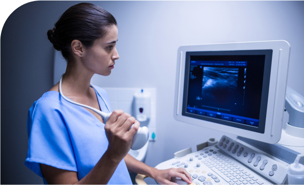
Ultrasound (US or ultrasonography) is much of the time the first imaging test done in quite a while who have side effects like jaundice or torment in the right upper piece of their midsection. The doctor might choose the next best investigation based on the ultrasound’s results.
A CT filter is a sort of x-beam that gives an image of organs and different designs (counting any cancers) in your body. It is used to examine a cancer in greater detail and its relationship to your body’s surrounding organs. It likewise gives data connected with disease spread into the lymph hubs, liver or lungs.

Endoscopic ultrasound scan (EUS): Dr. Deepak G. Chhabra is a Mumbai gallbladder cancer specialist who uses a special endoscope with an ultrasound probe and a small needle at the end. For more in-depth information about the tumor’s local spread, the scope is inserted through the mouth into the food pipe known as the oesophagus and the first part of the small intestine.
When planning for surgery, the doctor may be able to tell if and how far a tumor has invaded the gallbladder wall with the help of ultrasound. The presence of enlarged lymph nodes nearby, which may indicate that cancer has spread, can be detected by ultrasound.
Additionally, it can be utilized to direct a needle into a suspicious node so that cells can be extracted (biopsied) and examined under a microscope.

Positron emission tomography (PET) scan This test is performed in conjunction with a computed tomography (CT) scan by injecting a radioactive substance into the body. The goal of this procedure is to bring to light any and all locations throughout the body where the tumor currently exists or has the potential to spread.
After an MRI or CT scan, additional information may be gathered through this test. All patients do not require a PET-CT scan. This scan’s necessity will be determined by your physician.

Although imaging tests may indicate the presence of bile duct cancer, the majority of the time, a sample of bile duct cells or tissue is taken (biopsied) and examined under a microscope to confirm the diagnosis.
However, a biopsy may not always be performed prior to surgery to remove the tumor in cases where gallbladder cancer is likely. Doctors often worry that cancer cells could spread by inserting a needle into the tumor or otherwise disturbing it without completely removing it.
In the case of imaging tests (ultrasound, CT or X-ray examines, cholangiography, and so on.) if there are no obvious signs of distant spread and there is a gallbladder tumor, the doctor may decide to treat it as a gallbladder cancer with surgery right away. See the “Surgery” section.) After the gallbladder is removed in these instances, the tissue of the gallbladder is examined under a microscope.
A biopsy of a suspicious area in the gallbladder may be recommended by a doctor in other instances to confirm the diagnosis of gallbladder cancer. Imaging tests, for instance, may reveal that a tumor has grown too large or spread to be removed completely by surgery. Sadly, many gallbladder cancers cannot be removed when they are first discovered.
1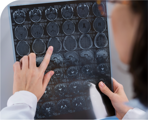
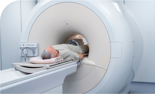
Similar to a CT scan, magnetic resonance imaging (MRI) creates images of your abdomen’s organs using magnetism rather than x-rays. Unlike a CT scan, an MRI does not cause any pain and the magnetism is harmless. In the event that the patient is allergic to the contrast dye injection that is used for CT scans, an MRI scan can be used to determine the extent of bile duct obstruction.
When examining the gallbladder, bile ducts, and other nearby organs, MRI scans can be very helpful because they provide a great deal of detail. They can sometimes assist in distinguishing a benign tumor from a malignant one.

In some cases, gallbladder cancer treatment may involve chemotherapy and/or radiation therapy, either alongside surgery or as an alternative if complete removal is not possible.
Dr. Deepak Chhabra, a leading gallbladder cancer surgeon in Mumbai, utilises advanced surgical techniques to enhance patient safety and outcomes. With improved medical care, post-operative monitoring, and modern anesthesia methods, gallbladder cancer surgeries have become safer and more effective.
For gallbladder cancer, there are two general types of surgical treatment: potentially curative surgery and palliative surgery.
When imaging tests indicate a good chance that the surgeon will be able to remove all of the cancer, potentially curative surgery is performed. The terms “resectable” and “unresectable” are terms used by doctors to describe cancers that, according to them, can be completely removed (by potentially curative surgery) and “resectable” refers to cancers that, according to them, have spread too far or are in a difficult location to be completely removed by surgery. Sadly, just a little part of bile pipe malignant growths are resectable at the time they are first found.
The goal of palliative surgery may be to alleviate symptoms or treat (or even prevent) complications like bile duct obstruction. When the tumor is too large to be completely removed, this kind of surgery is done. Although palliative surgery is not expected to cure cancer, it can sometimes improve a patient’s quality of life and extend their lifespan.
When gallbladder cancer is suspected, the surgeon frequently performs a laparoscopy first. This is done to help figure out how big the cancer is and whether or not it can be cut out. The surgeon may be able to see areas of cancer that were missed by imaging tests with laparoscopy. A laparoscope, a long, bright tube, is inserted into the abdomen through a small cut during this procedure. The laparoscope is used by the doctor to examine the abdominal cavity for evidence of cancer spread. Laparoscopy can also be helpful in planning the surgery to remove the cancer if it can be resected.


A cholecystectomy is an operation to remove the gallbladder. Simple cholecystectomy is the term used to describe an operation in which only the gallbladder is removed. This procedure is frequently performed to remove the gallbladder for other conditions, such as gallstones, but it is not performed if gallbladder cancer is known or suspected; instead, a more extensive procedure is performed.
After a person has a cholecystectomy for another reason, such as gallstones, gallbladder cancers may be discovered accidentally. There may not be a need for additional surgery if the cancer is found to be completely eradicated and in a very early stage (T1a). More extensive surgery may be recommended if the cancer has spread beyond the gallbladder.
In most cases of gallbladder cancer, a more extensive procedure known as an extended (or radical) cholecystectomy is performed due to the possibility that the cancer will return if only the gallbladder is removed. This can be a complicated, involved procedure that should be performed by a surgeon who has experience treating gallbladder cancer.
The extent of the surgery is determined by the location of the cancer and its potential spread. An extended cholecystectomy essentially eliminates:
If the surgeon feels it is needed and the patient is healthy enough, the operation may also include one or more of the following:
Palliative therapy is treatment that is given to help control or reduce symptoms caused by advanced cancer. It is not meant to be a curative treatment. If the cancer has spread too far to be completely removed by surgery, doctors may focus on palliative operations, palliative radiation, and other palliative therapies. Because these cancers tend to advance quickly, doctors try to use palliative therapies that are less likely to affect a person’s quality of life, when possible.
Mumbai-based gallbladder cancer specialist Dr. Deepak G. Chhabra performs this palliative procedure by connecting a portion of the bile duct that was blocked by the tumor with another portion of the intestine. The surgeon may simply request that a plastic or expandable metal tube—known as a stent—be inserted through the bile duct to keep it open in the event that a bypass cannot be performed.

Chemotherapy may be used following surgery to try to lower the risk of the cancer returning for resectable gallbladder cancers. This is called adjuvant chemotherapy. In an effort to increase the likelihood that surgery will be successful, some doctors may use it prior to it for cancers that are not resectable.
Treatment with high-energy rays or particles that kill cancer cells is known as radiation therapy. Radiation therapy comes in various forms.However, radiation therapy has not been widely used to treat gallbladder cancers, and nearly nine out of ten doctors who treat gallbladder cancers will not include radiation in their treatment plans.

Dr. Deepak Chhabra is a consultant Surgical Oncologist with an extensive experience in cancer surgeries. He is has specialized in Hepato (Liver) -Biliary (Gallbladder) and Pancreatic Cancer Surgeries.

Take the first step towards your journey to wellness by scheduling an appointment with Dr. Deepak Chhabra, a trusted oncologist dedicated to providing compassionate care and personalized treatment plans.
Discover first hand accounts from patients who have experienced compassionate care and expert treatment at our clinic. Read their reviews to get to know their journey.
5 Out of 5 from 92 Reviews


 VIDEO CONSULTATION
 Phone Calls
 Physical visit