
Breast cancer can be challenging, often diagnosed at later stages. However, with the expertise of a leading breast cancer surgeon in Mumbai, early detection, advanced treatments, and dedicated support, we aim to improve outcomes and bring hope to those affected.

With extensive training, experience, and dedication to patient care offering the comprehensive services tailored to meet the unique needs of each individual.

A woman can develop a wide range of breast conditions over her lifetime. These include a variety of benign (noncancerous) lumps and the normal changes that occur during the menstrual cycle. The fact that they are not cancerous unites them. Approximately 80% of breast lumps that require a biopsy turn out to be benign.

Breast Cancer In today’s Indian women, breast cancer is the most prevalent type of cancer. Every year, more than 100,000 women are diagnosed with breast cancer, and most people are aware of at least one person who has undergone treatment for the disease.
The good news is that breast cancer is being found earlier, when the tumor is only in the breast. Two-thirds of newly diagnosed breast cancers do not appear to have spread beyond the breast at this time.

A woman’s risk of developing breast cancer is average because she is female and gets older. She is more likely to develop breast cancer as she gets older. The majority of cases of breast cancer in women over the age of 50 occur in women over the age of 50, and the risk is particularly high in women over the age of 60.
No lady ought to see herself as excessively old to require ordinary screening mammograms.
Delve into the intricate web of factors intertwined with breast cancer, encompassing genetic predispositions, hormonal intricacies, and lifestyle habits, each playing a crucial role in shaping the susceptibility to this prevalent malignancy.

Developing breast cancer increases if her female member had breast cancer, especially at a young age.

There is evidence to suggest that a woman is more likely to develop this if she is exposed to estrogen for longer time.

Delayed childbirth increases breast cancer risk compared to early childbirth.

Under 30s, especially those treated with Hodgkin’s radiation, face higher breast cancer risk later.

Studies suggest slightly higher breast cancer risk with alcohol consumption.

Breast cancer survivors are more likely to develop the disease in their other breast.


Most of the time, early breast cancer does not cause pain. In point of fact, there may be no symptoms at all when breast cancer first appears.
A woman should see her doctor about any symptoms like these. Most often, they are not cancer, but it’s important to check with the doctor so that any problems can be diagnosed and treated as early as possible.

Physical changes in the breast can include lumps or thickening, changes in size or shape, nipple discharge, or skin dimpling, which should be promptly evaluated by a healthcare provider.

Nipple abnormalities encompass a range of changes that may indicate underlying breast health concerns. These include nipple discharge, which can vary in color and consistency and may occur spontaneously or upon manipulation.

Skin alterations can manifest as changes in color, texture, or appearance such as new moles, itching, or skin that appears scaly or crusty, often indicating the need for medical assessment.

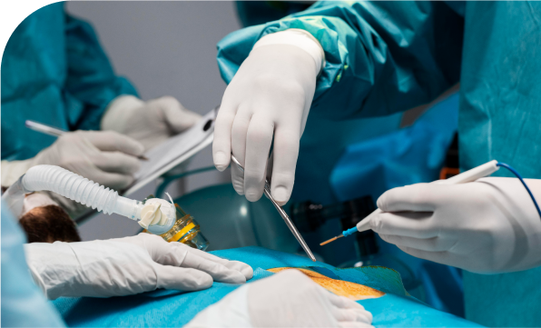
Breast cancer treatment in Mumbai focuses on removing early-stage cancer and reducing the risk of it spreading or returning. Treatment options may include surgery, radiation, chemotherapy, and hormone therapy, personalised to each patient’s needs.
Regular Breast Self-Examination (BSE) is vital for women 20+, done monthly post-period or on a set day post-menopause. It’s free, quick, and detects smaller tumors. Any change needs immediate attention. Early detection is vital against breast cancer.
Breast examination on a clinical basis: By palpating the lump and surrounding tissue, doctors distinguish between benign and cancerous lumps based on size, texture, and mobility. Armpit examination for abnormal lymph nodes is also conducted.
Mammography : A mammogram, a low-dose X-ray of the breast, is a crucial screening tool for early breast cancer detection, enhancing treatment success and survival rates. Scheduling the mammogram when breasts are least tender, usually after the period, can reduce discomfort. Annual screening mammograms are recommended from age 50. While mammograms may not detect all types of lumps, they become more effective with age as breasts become less dense.

Using high-frequency sound waves, ultrasonography can often show whether a lump is a fluid-filled cyst (not cancer) or a solid mass (which may or may not be cancer).This exam may be used along with mammography and is called Sonomammography. Based on these exams, the doctor may decide that no further tests are needed and no treatment is necessary. In such cases the doctor may need to check your breasts more regularly to watch for any changes.

A thin needle is used to take some cells from the breast lump or abnormal area. Sometimes an ultrasound is used to help guide the needle. The test is a similar to having blood taken for a blood test.It is usually done in a specialist’s rooms, a hospital outpatient department or at a radiology practice.

A wider needle is used to remove a small piece of tissue, called a core, from the lump or abnormal area. It is usually done under local anaesthetic. A mammogram or ultrasound is used to help guide the needle.

If the lump is too small to be biopsied using the method above, a surgical biopsy is needed. A surgical biopsy involves the surgical removal of a sample of tissue from a suspicious area in the body for examination under a microscope to determine if it is cancerous or benign.
Sometimes mammography may detect abnormalities in the breast (micro calcifications) without any lump in the breast. This may need to be tested. To help the surgeon find the abnormal tissue, a needle and wire may be put into the breast with the help of sonography guidance just before the biopsy. The biopsy is then done in a separate operation using a general anaesthetic. The lump and a small area of normal breast tissue around the lump are removed, along with the wire. This operation is usually done as day surgery but may mean an overnight stay in hospital. If the surgical biopsy removes all the cancer, further surgery may not be needed.

Your doctor will suggest a biopsy if an abnormal or unusual area of tissue is found in your breast. You may need one or more biopsies. A biopsy removes a small amount of breast tissue. There are a few ways of doing this. After a biopsy a pathologist examines the removed tissue.

If your breast cancer hasn’t spread, surgery is often the primary treatment option. Dr. Deepak Chhabra, a renowned breast cancer surgeon in Mumbai, specialises in advanced surgical procedures to remove tumours and minimise the risk of recurrence. Even in cases where the cancer has spread, his expertise ensures the best possible treatment approach for improved outcomes.

As the name suggests, the breast is not removed but conserved while only the abnormal lump is removed. Breast conserving surgery is offered if the cancer is small compared to the size of your breast. The surgeon removes the entire abnormal lump along with a sufficient margin of surrounding normal breast tissue (Wide local excision). This is then sent for testing immediately (Frozen section studies) to determine if all the abnormal tissue from the breast has been removed. The doctor gets the result within 10 minutes.
Radiotherapy (radiation treatment) is a must to the breast after a breast conserving surgery and is a part of Breast Conservation treatment.
Surgery to remove the whole breast is called mastectomy. The nipple and the surrounding dark area (areola) is also removed. The chest muscles are not removed. Some or all of the lymph nodes in the armpit closest to your affected breast may also be removed. You may be offered a mastectomy if the cancer is large compared to the size of the breast or the cancer is in more than one area of the breast.

After an anaesthetic, the movement of the bowel slows down and usually takes about 72 hours to get back to normal. After about 48-72 hours you will probably be ready to start taking small sips of water, however your doctors will tell you when it is appropriate for you to start drinking some fluids. This will be gradually increased after a couple of days until you are able to eat a light diet.
You will probably be ready to go home in about 10-14 days after your operation and once your stitches have been removed. If deemed appropriate your doctor may send you home with stitches and call you later to remove the stitches. By and large you should be able to climb several flights of stairs after your discharge from the hospital and you will be given diet instructions.
Before you leave hospital you will be given an appointment for a post-operative check-up at the outpatient clinic.
After mastectomy or armpit surgery, regular exercises are crucial for recovery. Aim to regain full arm and shoulder movement, reduce stiffness and swelling, and manage pain. Begin with gentle exercises like shoulder circles and basic arm movements, gradually increasing intensity as you regain strength and confidence.

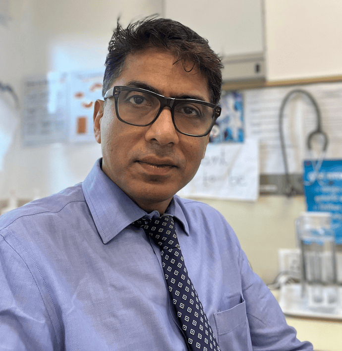
Dr. Deepak Chhabra is a consultant Surgical Oncologist with an extensive experience in cancer surgeries. He is has specialized in Hepato (Liver) -Biliary (Gallbladder) and Pancreatic Cancer Surgeries.
Take the first step towards your journey to wellness by scheduling an appointment with Dr. Deepak Chhabra, a trusted oncologist dedicated to providing compassionate care and personalized treatment plans.
Discover first hand accounts from patients who have experienced compassionate care and expert treatment at our clinic. Read their reviews to get to know their journey.
5 Out of 5 from 92 Reviews


 VIDEO CONSULTATION
 Phone Calls
 Physical visit