

With extensive experience, advanced treatments, and a deep commitment to patient care, we provide bile duct cancer treatment in Mumbai (India) with hope and healing at its core. At every step, we strive to offer personalised care, innovative solutions, and unwavering support to help patients move toward a healthier future.

The bile duct is a thin tube, about 4 to 5 inches long, that reaches from the liver to the small intestine. The major function of the bile duct is to move a fluid called bile from the liver and gallbladder to the small intestine, where it helps digest the fats in foods.

Bile duct cancers are commonly called CHOLANGIOCARCINOMAs.Cancers can develop in any part of the bile duct and, based on their location (see picture below), are divided into 4 groups:
These cancers develop in the smaller bile duct branches inside the liver. They can sometimes be confused with cancers that start in the liver cells, which are called hepatocellular carcinomas, and are often treated the same way. Only about 1 out of 10 bile duct cancers are intrahepatic.
These cancers develop at the hilum – where the hepatic ducts have joined and are just leaving the liver. They are also called “Klatskin tumors”. These are the most common type of bile duct cancer.
These cancers are rare and are found at the mid level of bile duct. More often than not these cancers are a part of cancers of the Gallbladder involving the bile duct.
These bile duct cancers are found further down the bile duct, closer to the small intestine. Because these bile ducts are outside of the liver, these cancers are also known as extrahepatic bile duct cancers.
More than 95% of bile duct cancers are of the adenocarcinoma type. Adenocarcinomas are cancers of glandular cells that can develop in several organs of the body. Bile duct adenocarcinomas develop from the mucus glands that line the inside of the duct. Cholangiocarcinoma is another name for a bile duct adenocarcinoma.
The exact causes of bile duct cancers is not know. But some factors can increase your risk of developing them. But risk factors don’t tell us everything. Having a risk factor, or even several risk factors, does not necessarily mean that a person will get the disease. And many people who get the disease may not have had any known risk factors.
In some Asian countries, infection by liver flukes (which are tiny parasite worms) can occur by eating poorly cooked fish. In humans, these flukes live in the bile ducts and can cause bile duct cancer. There are several types of liver flukes. The ones most closely related to bile duct cancer risk are called Clonorchis sinensis and Opisthorchis viverrini.
Being overweight or obese can increase the risk of developing cancers of the gallbladder and bile ducts. This may be because obesity increases the risk of gallstones and bile duct stones.
A risk factor is anything that affects your chance of getting a disease like cancer. Different cancers have different risk factors. Smoking is a risk factor for cancers of the lung, larynx , colon, bladder, kidney, and many other organs.

Signs and symptoms may not be present until the later stages of bile duct cancer, but in some cases they may lead to an early diagnosis. If you go to your doctor when you first notice symptoms, your cancer might be diagnosed at an early stage, when it is most treatable.
When bile duct cancer does cause symptoms, it is usually because the bile duct is blocked.
These are symptoms and signs of bile duct cancer, but it is important to remember that they are more likely to be caused by non-cancerous diseases. For example, people with gallstones may have many of these same symptoms. There are many causes of abdominal pain that are far more common than bile duct cancer. And hepatitis (inflamed liver most often caused by infection with a virus) is a much more common cause of jaundice. Still, if you have any of these problems, it’s important to see your doctor right away so the cause can be found and treated, if needed.

Symptoms may include persistent fever, nausea, and vomiting, which can indicate bile duct cancer and should prompt medical evaluation for proper diagnosis and treatment.

Jaundice, a yellowing of the skin and eyes, can occur when gallbladder cancer blocks the bile duct, preventing bile drainage. This buildup of bilirubin in the blood causes the discoloration. Some patients may experience jaundice.

Itching, especially in the absence of a rash, can be a symptom of bile duct cancer due to bile buildup in the bloodstream, requiring medical assessment to determine the cause and appropriate management.
Jaundice is yellowing of the skin and eyes that is caused by the build-up of a substance called bilirubin. This is the most common symptom of bile duct cancer.
It is important to realize that most cases of jaundice are not caused by cancer. It is more often due to hepatitis (inflammation of the liver) or a gallstone that has traveled to the bile duct. But whenever jaundice occurs, a doctor should be seen right away.


Excess bilirubin in the blood can also reach the skin, which can cause itching. Most people with bile duct cancer notice itching.

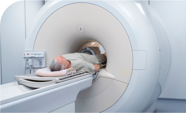
This section lists common tests that may be performed and it is not necessary for all the tests to be performed for a patient. Your doctor will select the tests that will assist him /her to have the maximum information about the tumor/ disease.
Tests of liver and gallbladder function: The doctor may order lab tests to find out how much bilirubin is in the blood. Bilirubin is the chemical that causes jaundice. Problems in the bile duct, gallbladder, or liver may cause too much bilirubin to remain in the blood. A high bilirubin count tells the doctor that there may be problems with the bile duct, gallbladder, or liver.
The doctor may also order tests for other substances in your blood, such as albumin, alkaline phosphatase, AST, ALT, and GGT. These are sometimes called liver enzymes or liver function tests. They can also give an indication of bile duct, gallbladder, or liver disease. Higher levels of these substances may point to blockage of the bile duct, but they cannot show if it is due to cancer or some other reason.

Ultrasound (US or ultrasonography) is much of the time the first imaging test done in quite a while who have side effects like jaundice or torment in the right upper piece of their midsection. The doctor might choose the next best investigation based on the ultrasound’s results.
A CT scan is a type of x-ray that gives a picture of organs and other structures (including any tumours) in your body. It is used to see more details of a cancer and its relation to the surrounding organs in your body. It also gives information related to cancer spread into the lymph nodes, liver or lungs.

This involves a special endoscope equipped with an ultrasound probe and a small needle at the end. The scope is placed through the mouth into the oesophagus (food pipe) and the first portion of the small intestine for more detailed information about the local spread of the tumor. EUS also allows the physician to get a tissue sample (biopsy) of the tumor.

This test may be used to build up more information after an MRI or CT scan. PET-CT scan is not necessary for all patients. Your doctor will decide if you need to undergo this scan.
This test is combined with a CT scan by injecting a radioactive material in the body to highlight all areas where the tumor has or can spread.

Imaging tests can suggest that a bile duct cancer is likely to be present, but in many cases a sample of bile duct cells or tissue is removed (biopsied) and looked at under a microscope to be sure of the diagnosis.
But a biopsy may not always be done before surgery for a possible bile duct cancer. If imaging tests (ultrasound, CT or MRI scans, cholangiography, etc.) suggest there is a tumor in the bile duct, the doctor may decide to proceed directly to surgery and to treat it as a bile duct cancer.
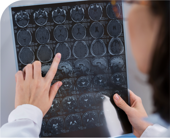
This test is like a CT scan, but it uses magnetism instead of x-rays to build up pictures of the organs in your abdomen. Like a CT scan, MRI is painless and the magnetism is harmless. MRI scan may be used to see the extent of blockage of bile duct and in case the patient is allergic to contrast dye injection used for CT scans.
MRI scans provide a great deal of detail and can be very helpful in looking at the bile ducts and nearby organs. Sometimes they can help tell a benign tumor from a malignant one.
Special types of MRI scans called MR cholangiopancreatography (MRCP) are used in people who may have bile duct cancer.

This is another way for your doctor to look at your bile duct. In this procedure, the doctor places a thin, hollow needle through the skin and into a bile duct within the liver. (A local anesthetic is used to numb the area before inserting the needle.)
A contrast dye is then injected through the needle, and x-rays are taken as it passes through the bile ducts. Like ERCP, this approach can also be used to take samples of fluid or tissues or to place stents (small, hollow tubes) in the bile duct to help keep it open.

In this test, a thin, flexible ‘telescope’ called an endoscope is put into your mouth then passed down your throat into your digestive system so that the doctor can examine you inside. The procedure is called ERCP, or endoscopic retrograde cholangio-pancreatography. Through the endoscope, the doctor can inject a liquid directly into the pancreatic duct and bile duct, allowing images of these organs to appear on x-ray pictures.
The test can show blockages and inflammation in these ducts, and allow the doctor to judge whether these are due to cancer or other problems. The doctor is also able to take a tissue or fluid sample through the endoscope, to help with the diagnosis. It can also be used to place a stent (a small tube) into a duct to help keep it open.

Tumor markers are substances made by cancer cells that can sometimes be found in the blood. People with bile duct cancer may have high blood levels of the carcinoembryonic antigen (CEA) and CA 19-9 tumor markers. High amounts of these substances often mean that cancer is present, but the high levels can be caused by problems other than bile duct cancers.
Also, not all bile duct cancers make these tumor markers, so low or normal levels do not mean there is no cancer.
There are 2 general types of surgical treatment for bile duct cancer — potentially curative surgery and palliative surgery.
This surgery is used when imaging tests indicate a good chance that the surgeon will be able to remove all of the cancer. Doctors may use the term resectable to describe cancers they believe can be removed completely (by potentially curative surgery) and unresectable to describe those they think have spread too far or are in too difficult a place to be entirely removed by surgery. Unfortunately, only a small portion of bile duct cancers are resectable at the time they are first found.
Palliative surgery may be performed to relieve symptoms or treat (or even prevent) complications, such as blockage of the bile ducts. This type of surgery is performed when the tumor is too widespread to be completely removed. Palliative surgery is not expected to cure the cancer, but it can sometimes help someone feel better and sometimes can even help them live longer.

For resectable cancers, the type of operation depends on the location of the cancer.
These cancers have started in bile ducts within the liver. To treat these cancers, the surgeon cuts out the part of the liver containing the cancer. Removing part of the liver is called a partial hepatectomy. Sometimes this means that a whole lobe of the liver must be removed. This is called hepatic lobectomy. As much as 70 percent of the liver may be removed if the remaining liver is healthy. The remaining healthy liver can then take care of the body functions. Also, the liver can re-grow some of the missing part. The new cells grow over several weeks.
These cancers begin where the branches of the bile duct first exit the liver. Surgery for these cancers requires great skill, as the operation is quite extensive. Usually part of the liver must be removed along with the entire bile duct, gallbladder and nearby lymph nodes. Then the surgeon must connect the remaining ducts to the small intestine.
It becomes important for the patient to understand that a major portion of the liver needs to be removed as the bile duct system is connected to that lobe of the liver. So for the patient this is an extensive liver surgery
These cancers are further down the bile duct near the pancreas and small intestine. Along with the bile duct and nearby lymph nodes, in most cases the surgeon must remove part of the pancreas and small intestine. The procedure is same as that performed for a pancreatic cancer (Whipple’s operation). See treatment of pancreatic cancer.
It becomes important for the patient to understand that a major portion of the liver needs to be removed as the bile duct system is connected to that lobe of the liver. So for the patient this is an extensive liver surgery
Palliative therapy is treatment that is given to help control or reduce symptoms caused by advanced cancer. It is not meant to be a curative treatment. If the cancer has spread too far to be completely removed by surgery, doctors may focus on palliative operations, palliative radiation, and other palliative therapies. Because these cancers tend to advance quickly, doctors try to use palliative therapies that are less likely to affect a person’s quality of life, when possible.

For bile duct cancers that are resectable, chemotherapy may be used after surgery (often along with radiation therapy) to try to lower the risk that the cancer will return. This is known as adjuvant chemo. Some doctors may use it before surgery for borderline resectable cancers to try to improve the odds that surgery will be successful. This is called neoadjuvant treatment. Chemotherapy may also be used (with or without radiation therapy) for more advanced cancers. By and large there are very few responses to chemotherapy for Bile duct cancers.
Radiation therapy is treatment with high-energy rays or particles that destroy cancer cells. There are different kinds of radiation therapy.
Three-dimensional conformal radiation therapy (3D-CRT) uses special computers to precisely map the location of the tumor(s). Radiation beams are shaped and aimed at the tumor(s) from several directions, which makes it less likely to damage normal tissues. Most doctors now recommend using 3D-CRT when it is available.

Dr. Deepak Chhabra is a consultant Surgical Oncologist with an extensive experience in cancer surgeries. He is has specialized in Hepato (Liver) -Biliary (Gallbladder) and Pancreatic Cancer Surgeries.

Take the first step towards your journey to wellness by scheduling an appointment with Dr. Deepak Chhabra, a trusted oncologist dedicated to providing compassionate care and personalized treatment plans, offering advanced bile duct cancer treatment in Mumbai (India).
Discover first hand accounts from patients who have experienced compassionate care and expert treatment at our clinic. Read their reviews to get to know their journey.
5 Out of 5 from 92 Reviews


 VIDEO CONSULTATION
 Phone Calls
 Physical visit